This activity investigates the role of sleep on brain function, by considering the results of an experiment performed on mice.
Background Information
Nearly all animals require sleep, though the reasons for this are not fully understood. However, the fact that sleep has been evolutionarily conserved suggests that it serves vital biological functions. Lack of sleep impairs brain function and, in prolonged cases, can cause dementia or death. Surprisingly, most of the mechanisms by which sleep preserves brain function remain a mystery. In this study, scientists set out to investigate whether sleep plays a role in removing metabolites (the molecules produced during normal metabolism) from the brain. Metabolites can impair neurological function when they accumulate at abnormally high levels. One such metabolite is a protein called β-amyloid (Aβ). During cell metabolism, it is deposited in the spaces between brain cells (called interstitial spaces) and then removed by circulating cerebrospinal fluid. The buildup of Aβ proteins in the brain is linked to neurodegenerative diseases like Alzheimer’s disease (AD).
Previous research has shown that levels of Aβ are higher in the brains of animals that are awake than in those that are asleep, so the researchers in this study tested whether the rate of Aβ removal is higher during sleep. The scientists injected radioactively labeled Aβ into the brains of 25 awake mice, 29 that were sleeping naturally, and 23 that were anesthetized via a thin tube previously installed in the skull. The anesthetized mouse group was used to determine whether differences in Aβ clearance rates that may be seen between asleep and awake states are due to circadian rhythms, which do not occur under anesthesia. At several time points between 10 and 240 minutes post-injection, three to six mice per time point were humanely euthanized in order to measure the levels of labeled Aβ in their brains and determine the clearance rate.
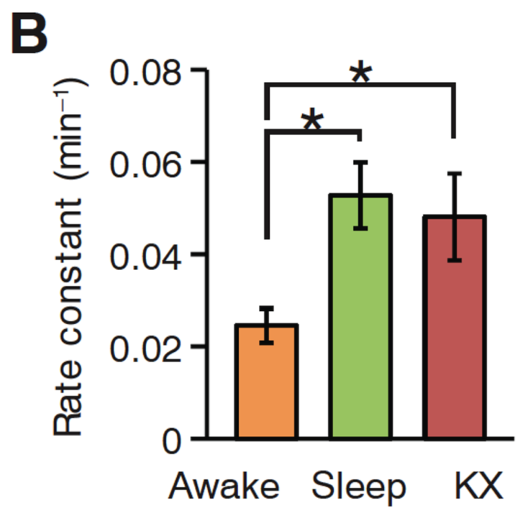
Caption: The different rates at which β-amyloid (Aβ) was cleared from the brains of mice that were awake, asleep, or in an induced sleep state using the anesthetic ketamine/xylazine (KX). The * indicates a P value < 0.05, meaning that the differences in clearance rates are statistically significant in this study. The error bars represent standard error of the mean.
Discussion Questions
- Rate is expressed as change over time. Describe what change is being studied in this experiment. (Understanding the x- and y-axes should be helpful)
- How would you describe the difference between awake and sleeping mice in terms of Aβ clearance rate?
- Does the data in this figure support the researchers’ hypothesis? In what ways?
- Is there a significant difference in the rate of Aβ clearance between sleeping and anesthetized mice? How do you know? How would you interpret the error bars in this figure?
- What is the purpose of including anesthetized (induced sleep) mice in this experiment?
- Why did the scientists use radioactively labeled Aβ instead of unlabeled Aβ to measure Aβ clearance int he mouse brains? What drawbacks/limitations might there be in using injected, radio labeled Aβ as a proxy for natural sources of Aβ in the brain?
- Based on the data in this figure, why do you think that people with insomnia have decreased brain function?
- Why do you think the scientists chose to use mice as a model for brain function and sleep?
SOURCE
L.Xie et al. 2013. Sleep drives metabolite clearance from the adult brain. Science 342 (6156), 373-377. View article: http://science.sciencemag.org/content/342/6156/373
AUTHOR: Natalie Dutrow, PhD, Judge Memorial Catholic High School, Salt Lake City, UT. Edited by: Hilary Gerstein, PhD, Center for Neuroscience & Society, University of Pennsylvania; Satoshi Amagai, PhD, Bridget Conneely, Melissa Csikari, and Jessica Johnson, HHMI