In the Virtual Microscope app, locate the Bacterial Slides in the Slide Catalog. View each slide listed up to the 10X, or 40X objective as needed. Hint: You may have to drag the slide around to get a good view of the specimens. Describe what you see and, where prompted, sketch the specimens in the charts below. In particular, think about how the shapes differ between the types of bacteria, and how the cells are arranged with respect to one another.
Acid Fast Mix
The acid–fast stain is a differential stain used to identify acid–fast organisms such as members of the genus Mycobacterium . Acid–fast organisms are characterized by wax-like, nearly impermeable cell walls; they contain mycolic acid and large amounts of fatty acids, waxes, and complex lipids.
1. What two types of bacteria are included on this slide?
2. Describe what you see at 400x magnification
3. Now, perform an oil immersion. Describe any details that are visible now at 1000x, that you could not see at 400x.
4. View the links below. Now, is it easier to understand what you were viewing on the slide?
Click here to view M. smegmatis under even higher magnification: https://en.wikipedia.org/wiki/Mycobacterium_smegmatis#/media/File:Mycobacterium_smegmatis.tif
Click here to view S. aureus under higher magnification: https://en.wikipedia.org/wiki/Staphylococcus_aureus
Endospore Stain
The endospore stain is a differential stain used to visualize bacterial endospores. Endospores are formed by a few genera of bacteria, such as Bacillus . By forming spores, bacteria can survive in hostile conditions. Spores are resistant to heat, dessication, chemicals, and radiation.
1. What type of bacteria is included on this slide?
2. Work up through the objective lens, and sketch what you see at every step.
3. Describe what you see at 400x magnification
4. Now, perform an oil immersion. Describe any details that are visible now at 1000x, that you could not see at 400x.
5. View this link (https://www.austincc.edu/microbugz/endospore_stain.php). Now, is it easier to understand what you were viewing on the slide?
Gram Stain
Gram staining is a common technique used to differentiate two large groups of bacteria based on their different cell wall constituents. The Gram stain procedure distinguishes between Gram positive and Gram negative groups by coloring these cells red or violet. Gram positive bacteria stain violet due to the presence of a thick layer of peptidoglycan in their cell walls, which retains the crystal violet these cells are stained with. Alternatively, Gram negative bacteria stain red, which is attributed to a thinner peptidoglycan wall, which does not retain the crystal violet during the decoloring process.
1. What two types of bacteria are included on this slide? Which appears to be the gram negative strain? Which is the gram positive? How do you know?
2. Work up through the objective lens, and sketch what you see at every step.
3. Describe what you see at 400x magnification
4. Now, perform an oil immersion. Describe any details that are visible now at 1000x, that you could not see at 400x.
Bacterial Shapes
Now, we are going to put away the virtual microscope, and view some images to get a better idea of prokaryote diversity. In the images below, observe the three basic shapes of bacteria (refer to your text book, in the chapter on prokaryotes). Look for single cells that are isolated from the more obvious, large mass of cells. Draw and label each form in the space below.
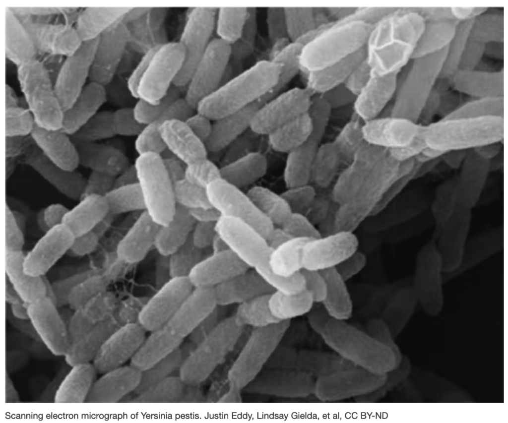
Bubonic Plague 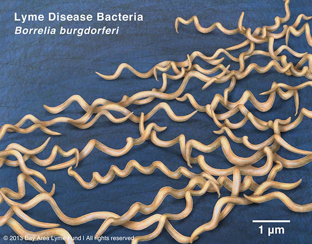
Lyme Disease 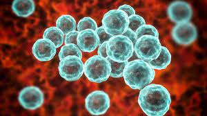
Chlamydia

1. What disease is caused by B. burgdorferi?
Cyanobacteria
Cyanobacteria are larger than many other prokaryote species and were formerly called blue-green algae because of their ability to carry out photosynthesis. These organisms live in many environments, including desert soils – where they become active when they encounter moisture –and various oceanic habitats.
Anabaena: Cells of this cyanobacterium are found in filaments, which contain specialized cells known as heterocysts.
- View the video of an Anabaena culture and carefully draw the filaments. Label the heterocysts and any other structures you see.
- What is the function of the heterocysts?
Nostoc: Cells of this cyanobacterium are also found in filaments and also possess heterocysts.
- Draw filaments of Nostoc and label the heterocysts.
Oscillatoria: Cells of this cyanobacterium are also found in filaments.
- Compare the appearance of Oscillatoria to the other two cyanobacteria
- Describe any movements that you see
- Speculate as to the origin of its name.
Archaean Diversity
Until relatively recently, prokaryotes were regarded as a single group of organisms, classified based on the structures of their cell walls, their shapes, and the substances they consume. In 1965, it was proposed that they be classified using gene sequences, and this phylogenetic approach is the main method used today.
In 1977, Archaea were classified separately for the first time, based on their ribosomal RNA (rRNA) genes. The first evidence for Archaebacteria as a separate line of descent included the lack of peptidoglycan in their cell walls, two unusual coenzymes, and the results of 16S ribosomal RNA gene sequencing. This led to the reclassification of known organisms into three natural domains: Eukarya, Bacteria and Archaea.
The word archaea means “ancient.” The first described representatives were methanogens, assumed to have a metabolism that reflected Earth’s primitive atmosphere. Later, extreme halophilic and hyperthermophilic microbes were also included in Archaea. For a long time, archaea were seen as extremophiles that exist only in extreme habitats such as hot springs and salt lakes, but by the end of the 20th century, archaea had been identified in non-extreme environments as well. Today, they are known to be a large and diverse group of organisms abundantly distributed throughout nature.
This video has more information, including interviews, with the man who “discovered” the Archaea.
- Who first realized that the Archaea should not be classified as bacteria?
- What is an extremophile?
- Where do halophiles live? What about thermophiles?