Activity: Working magnification
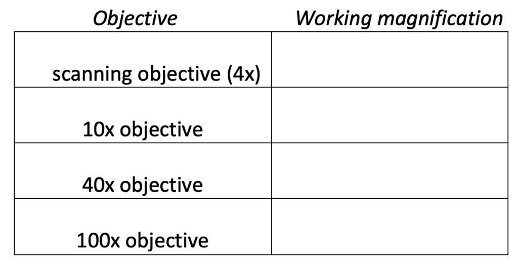
Working magnification (total magnification) is the product of the ocular magnification (usually 10x) and the objective lens magnification (ranging between 4x and 100x). The following terms refer to characteristics of a particular working magnification.
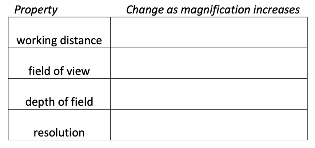
- Working distance is the distance between the focal plane (the plane in the specimen where you are focused) and the front of the lens.
- Field of view is the area in the focal plane that is visible through the objective.
- Depth of field is essentially the thickness of the focal plane, or more precisely, the range of depth in which the specimen is close enough to being in perfect focus.
- Resolution is the size of the smallest feature that can be distinguished.
Questions:
- Determine the working magnification for the following objectives:
2. How do the properties of working magnification change when you increase magnification?
Activity: Virtual Microscope
In this activity, we will explore a virtual microscope that is very similar to the way a traditional microscope works. You will learn the basics of light microscopy, comparable to what you would learn in a seated lab. You will identify components of the microscope, understand the functions of those components, learn how to focus in on a specimen, and review proper care and maintenance.
If you are working with other students, make sure that every student does the activity independently. We want everyone to get the experience with the microscope’s functions.
Open the virtual microscope here: http://www.ncbionetwork.org/iet/microscope/
“Learn” tab:
- Locate the Eyepiece Lens. Describe the purpose of this part of the microscope.
- Locate the Objective Lens. How do the different objective lenses differ from one another? From the Eyepiece Lens?
- Locate the Coarse Focus. What is the purpose of this knob?
- Now locate the Fine Focus. How does this knob differ from the Coarse Focus? Which of the two should you typically use first?
- Locate the Lens Paper, and Kimwipes. Which of these should you use to clean the microscope lenses? Which should you never use to clean lenses, and why?
- Which part of the microscope can be adjusted to change the amount of light passing through the stage opening?
- Which part of the microscope moves up and down, and forward and back?
“Explore” Link:
- What happened to the brightness of your specimen? Did you need to adjust the lighting? Why do you think this happened?
- Now click on the 100x lens. What happened? Why would you not want to skip lenses on an actual microscope?
- Select “Whitefish Metaphase.” Focus on the slide, and raise the magnification until you are viewing at 40x. Can you locate one of the cells that is in metaphase? Describe what you see.
- Now, select “Whitefish Telophase/Cytokinesis,” and view with the 40x lens. Can you locate one of the cells that is in cytokinesis? Describe what you see.
Activity: Focusing the Microscope
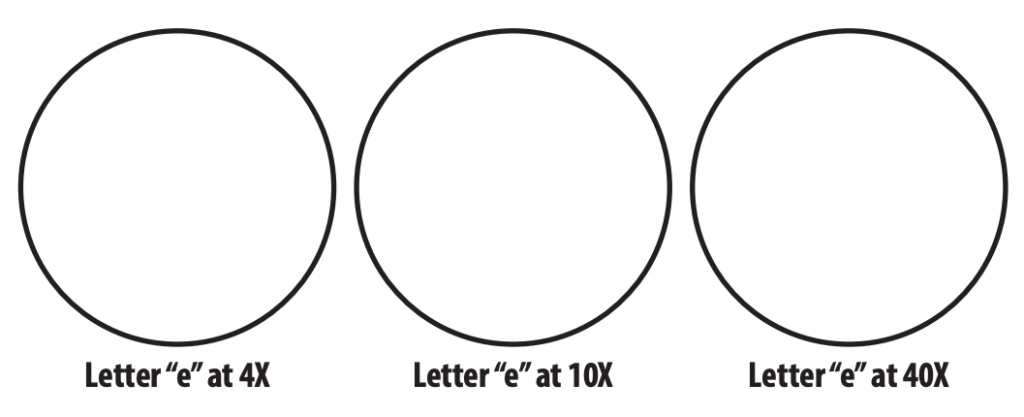
- Obtain the slide with the letter “e,” and focus it under the 4x objective. Draw what you see in the space provided below.
- Focus the 10x lens on the “e.” Draw what you see. Then focus the 40x lens on the “e.” Draw what you see.
Activity: Oil Immersion
- From the Slide Catalog, select “Animals” and then choose any of the Whitefish slides, which show the various stages of cell division.
- Focus and move through the objective lenses as described above.
- Now select 100x. You will be prompted to place a drop of oil on the slide.
- USING ONLY THE FINE FOCUS KNOB, bring the slide into view. Draw what you see.
- When finished, you will need to clean the objective lens USING ONLY LENS PAPER before you will be able to remove the slide.
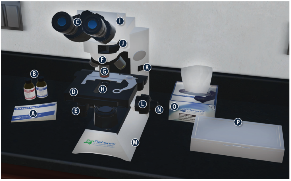
Activity: Review the Parts of the Microscope
Complete the chart below by matching the labels with the appropriate parts of the microscope.
