Ecdysozoa
The superphylum Ecdysozoa contains an incredibly large number of species, and is believed to be monophyletic—a clade consisting of all evolutionary descendants from one common ancestor. Ecdysozoa includes two of the most diverse animal groups: phylum Nematoda (the roundworms) and phylum Arthropoda (the arthropods). Tardigrada (“water bears”) are a relatively small phylum, but they have exceptional resistance to desiccation and other environmental hazards. The most prominent distinguishing feature of ecdysozoans is the cuticle—a tough but flexible exoskeleton that protects these animals from water loss, predators, and other dangers of the external environment. All members of this superphylum periodically go through a molting process that culminates in ecdysis—the actual shedding of the old exoskeleton. (The term “ecdysis” translates roughly as “take off” or “strip.”) During the molting process, old cuticle is replaced by a new cuticle, which is secreted beneath it, and which will last until the next growth period.
Phylum Nematoda
The name Nematoda is derived from the Greek word “Nemos,” which means “thread,” and includes all true roundworms. Nematodes are present in all habitats, typically with each species occurring in great abundance. The free-living nematode, Caenorhabditis elegans, has been extensively used as a model system for many different avenues of biological inquiry in laboratories all over the world.
The Nematoda, like other members of the superphylum Ecdysozoa, are triploblastic and possess an embryonic mesoderm that is sandwiched between the ectoderm and endoderm. They are also bilaterally symmetrical, meaning that a longitudinal section will divide them into right and left sides that are superficially symmetrical. In contrast with flatworms, nematodes are pseudocoelomates and show a tubular morphology and circular cross-section. Nematodes include both free-living and parasitic forms.
Morphology
These animals have a complete digestive system with a distinct mouth and anus, in contrast to only one opening in the digestive tract of flatworms. The mouth opens into a muscular pharynx and intestine, which leads to a rectum and anal opening at the posterior end. The epidermis can be either a single layer of cells or a syncytium—a multinucleated tissue that in this case is formed by the fusion of many single cells. The cuticle of nematodes is rich in collagen and a polymer called chitin, which forms a protective armor outside the epidermis. The cuticle extends into both ends of the digestive tract, the pharynx, and rectum. In the head, an anterior mouth opening is composed of three (or six) “lips” as well as teeth derived from the cuticle (in some species). Some nematodes may present other modifications of the cuticle such as rings, head shields, or warts. These external rings, however, do not reflect true internal body segmentation, which as we have seen is a hallmark of phylum Annelida. The attachment of the muscles of nematodes differs from that of most animals: they have a longitudinal layer only, and their direct attachment to the dorsal and ventral nerve cords creates a strong muscular contraction that results in a whiplike, almost spastic, body movement.
Excretory System
In nematodes, specialized excretory systems are not well developed. Nitrogenous wastes, largely in the form of ammonia, are released directly across the body wall. In some nematodes, osmoregulation and salt balance are performed by simple excretory cells or glands that may be connected to paired canals that release wastes through an anterior pore. In marine nematodes, the excretory cells are called renette cells, which are unique to nematodes.
Nervous system
Most nematodes have four longitudinal nerve cords that run along the length of the body in dorsal, ventral, and lateral positions. The ventral nerve cord is better developed than the dorsal and lateral cords. Nonetheless, all nerve cords fuse at the anterior end, to form a pharyngeal nerve ring around the pharynx, which acts as the head ganglion or the “brain” of the roundworm. A similar fusion forms a posterior ganglion at the tail. In C. elegans, the nervous system accounts for nearly one-third of the total number of cells in the animal!
Phylum Tardigrada
The tardigrades (“slow-steppers”) comprise a phylum of inconspicuous little animals living in marine, freshwater, or damp terrestrial environments throughout the world. They are commonly called “water bears” because of their plump bodies and the large claws on their stubby legs. There are over 1,000 species, most of which are less than 1 mm in length. A chitinous cuticle covers the body surface and may be divided into plates. Tardigrades are known for their ability to enter a state called cryptobiosis, which provides them with are resistance to multiple environmental challenges, including desiccation, very low temperatures, vacuum, high pressure, and radiation. They can suspend their metabolic activity for years, and survive the loss of up to 99% of their water content. Their remarkable resistance has recently been attributed to unique proteins that replace water in their cells and protect their internal cell structure and their DNA from damage.
Morphology and Physiology
Tardigrades have cylindrical bodies, with four pairs of legs terminating in a number of claws. The cuticle is periodically shed, including the cuticular covering of the claws. The first three pairs of legs are used for walking, and the posterior pair for clinging to the substrate. A circular mouth leads to a muscular pharynx and salivary glands. Tardigrades feed on plants, algae, or small animals. Plant cells are pierced with a chitinous stylet and the cellular contents are then sucked into the gut by the muscular pharynx. Bands of single muscle cells are attached to the various points of the epidermis and extend into the legs to provide ambulatory movement. The major body cavity is a hemocoel, but there are no specialized circulatory structures for moving the blood, nor are there specialized respiratory structures. Malpighian tubules in the hemocoel remove metabolic wastes and transport them to the gut. A dorsal brain is connected to a ventral nerve cord with segmental ganglia associated with the appendages. Sensory structures are greatly reduced, but there is a pair of simple eyespots on the head, and sensory cilia or bristles concentrated toward the head end of the animal.
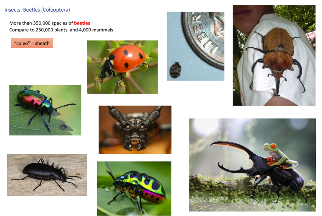
Phylum Arthropoda
The phylum Arthropoda is the most diverse and widespread group of animals on Earth. Arthropods are abundant in terrestrial, marine, and freshwater aquatic habitats. They make up over three-‐fourths of all currently known living and fossil animals, or over one million species in all. Since many arthropod species remain undocumented or undiscovered, especially in tropical rain forests, the true number of living arthropod species is probably in the tens of millions. One recent conservative estimate puts the number of arthropod species in tropical forests at 6 to 9 million species (Thomas, 1990).
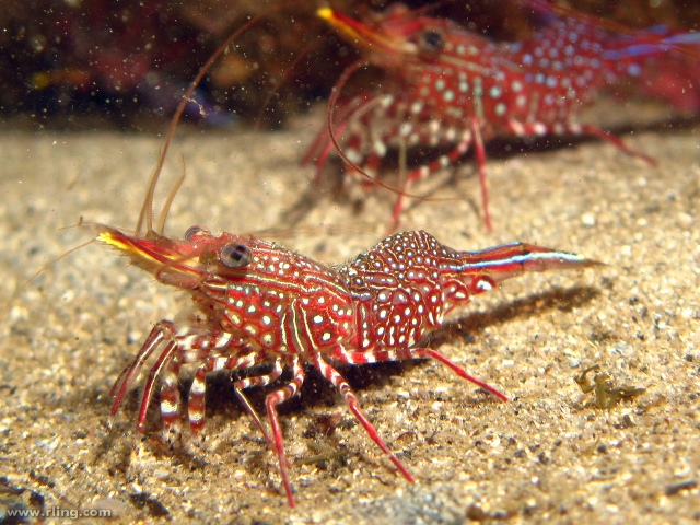
There are four main lineages of arthropods. The Trilobita, the first major adaptive radiation of arthropods, went extinct around 250 million years ago, but are known from an extensive fossil record. The Crustacea (crabs, shrimp and lobsters, and their relatives) usually inhabit marine or aquatic habitats. They have mandibles for feeding and they have biramous (2-‐branched) appendages. The Chelicerata (spiders, scorpions, mites, and horseshoe crabs) are marine or terrestrial. They have piercing mouthparts called chelicerae for feeding and book gills or book lungs for respiration (book, because of page-‐like arrangement of lamellae -‐ structures used for gas exchange). The Uniramia are divided into two main taxa. The Myriapoda (centipedes and millipedes) are terrestrial, have many segments with fairly undifferentiated legs, and use a tracheal system for respiration. The Hexapoda (insects) have three body regions (head, thorax, and abdomen), with six legs attached to the thorax. They are terrestrial or aquatic, and they also respire with a tracheal system. Insects are the most diverse group of arthropods.
Arthropods have colonized terrestrial habitats several times. Indeed, the colonization of land has even occurred more than once among Crustaceans (terrestrial hermit crabs, sow bugs). The chelicerates, the insects, and myriapods have also colonized land.
External morphology: external skeleton consisting mostly of chitin (what is the chemistry of chitin?); segmented body where individual segments are often fused together (tagmatization); appendages are also segmented (jointed appendages); Appendages vary in structure and function, i.e. they are specialized to perform different functions (sensory, feeding, locomotion); Coelom is present but greatly reduced to spaces around the heart and gonads; a hemocoel (blood filled spaces) made up of sinuses, blood vessels & heart is present.
Internal structure and physiology: Ventral nerve cord present; dorsal cerebral ganglion (brain, for what it’s worth); open circulatory system; dorsal heart; excretion through a gland (in Crustacea) or malpighian tubules (Hexapoda); respiration through diverse mechanisms (Crustacea-‐ gills and over body surface, Hexapoda-‐ tracheal system, Arachnida-‐ book lungs).
Development: Protostome developmental pattern (i.e. spiral cleavage, coelom formed by splitting of mesoderm, determinate development).
Classification
- Subphylum Crustacea. Possess: Mandibles (used for feeding); biramous (“two-‐ branches”) appendages; two pairs of antennae. Mostly marine or aquatic. There are several classes of crustaceans; we only list three of them here.
- Class Branchiopoda (“gill-‐foot”). No appendages on abdomen. Thoracic leaf shaped appendages for gas exchange. Brine shrimp (Artemia sp.) and water fleas (Daphnia sp.) are important components of zooplankton.
- Class Maxillopoda (“jaw-‐foot”). Large maxillae (feeding appendages). This group is extremely diverse in habitat use. Most species are free-‐living, but these may be either sessile filter feeders (barnacles), or mobile planktonic crustaceans (copepods). Rhizocephalans are bizarre internal parasites of crabs. Copepods can also be external parasites of fish.
- Class Malocostraca (“soft-‐shell”). Head, thorax, and abdominal segments present. Head and thorax often fused together and covered with a carapace. Appendages present on abdomen. Compound eyes usually present. Includes shrimp, lobsters, crabs, crayfish, and isopods (sow bugs).
- Subphylum Chelicerata. Body consists of a cephalothorax (resulting from the fusion of the head and thorax) and an abdomen. No antennae. The first pair of appendages are the chelicerae, which they are used to catch and pierce their prey. The second pair of appendages are the pedipalps, which vary greatly among groups and which can be used in prey capture. Some are marine, and some are terrestrial.
- Class Merostomata. Horseshoe crabs. Marine organisms. Book gills for respiration. Dorsal side covered with a carapace.
- Class Pycnogonida. Sea Spiders. Marine organisms. Simple eyes present. Only a vestigial abdomen present. No respiratory or excretory system.
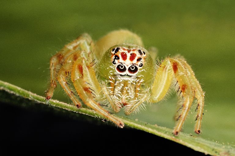
- Class Arachnida. Spiders, scorpions, pseudoscorpions, ticks, mites. Terrestrial. Waxy cuticle. Book lungs for respiration. Simple eyes present. 4 pairs of walking legs.
- Subphylum Myriapoda. Body divided into a head and a long trunk with many segments with legs on them. Relative lack of tagmatization (little fusion of segments). In contrast to the crustaceans, these organisms all have uniramous appendages. They are terrestrial
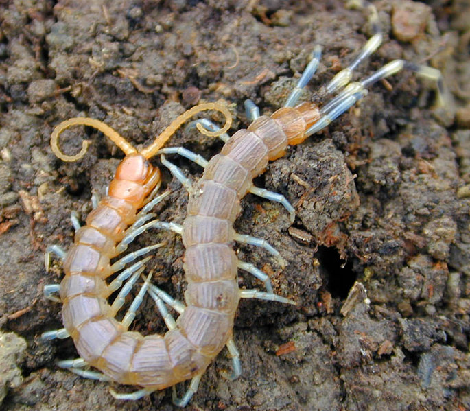
- Class Chilopoda. The centipedes. Flattened body with one pair of walking legs per segment. Appendages on first trunk segment modified from legs into poisonous fangs.
- Class Diplopoda. The millipedes. Two pair of walking legs per diplosegment (pairs of segments that appear to have fused), body often cylindrical.
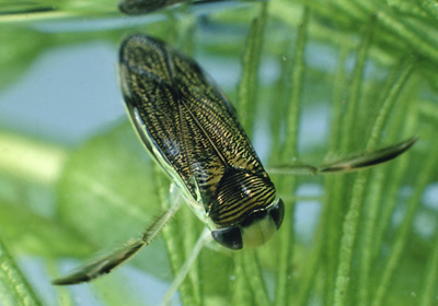
- Subphylum Hexapoda (Class Insecta). Terrestrial or aquatic (freshwater); adults often with wings. Body divided into three regions with at least some fused segments: head, thorax, and abdomen. Three pairs of segmented legs, one pair of antennae. They have a hardened exoskeleton. Compound eyes and mandibles for feeding.
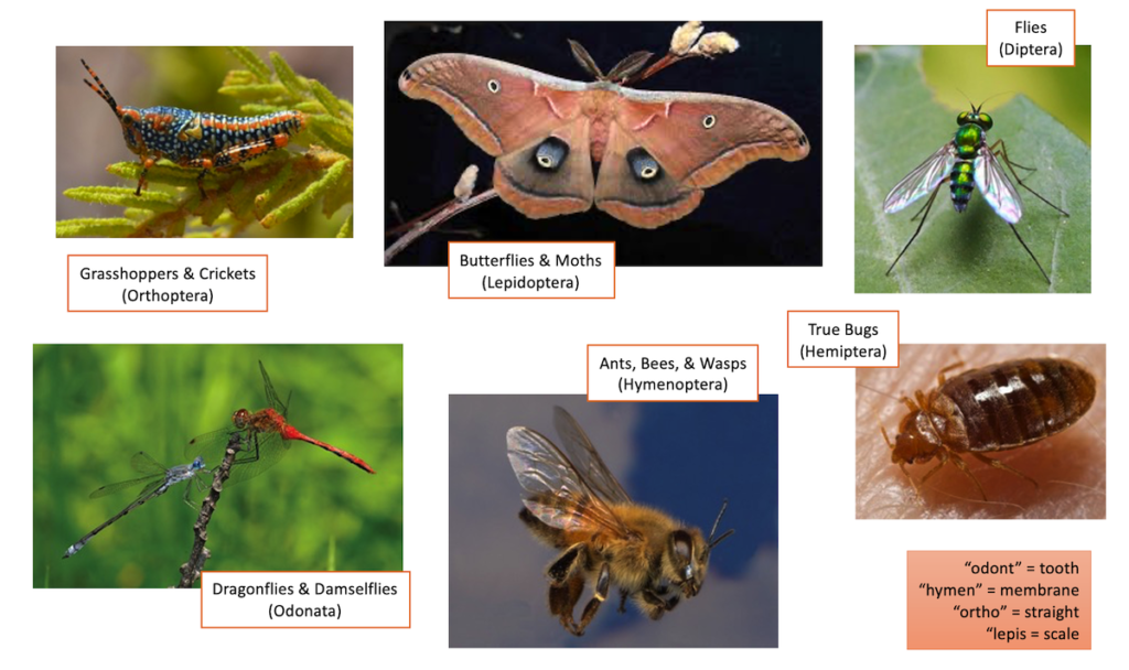
Major Orders of Insects:
Text adapted from Open Stax Biology 2e. Access for free at https://openstax.org/books/biology-2e/pages/1-introduction
Defensive Adaptations
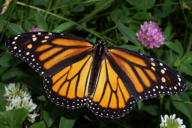
Evolutionary strategies that improve defense are common across a wide variety of organisms. Many of these strategies have one obvious purpose: to avoid being eaten by a predator. We will talk about two ways in which coloration and other aspects of morphology can provide this type of defense: aposematic coloration, and cryptic coloration.
Aposematic, or “warning” coloration is often expressed as bright, easily remembered patterns that signal danger to predators, discouraging them from attack. This coloration is often (but not always) accompanied by other traits that also discourage predation, such as bitter tasting compounds or toxins. For example, monarch butterflies taste bad and may cause vomiting in a predator who has eaten one. The first time a bird eats one, it is likely to learn that this was a mistake. The next time this bird sees a brightly-colored orange butterfly, it will avoid it because of this previous experience. Organisms that are venomous or poisonous – for example, dart frogs that release toxins from their skin – are also often brightly colored as a way of discouraging attacks. “Startle” coloration, such as a moth whose wings have a pattern that resembles the eyes of an owl, can be displayed to frighten away potential predators.
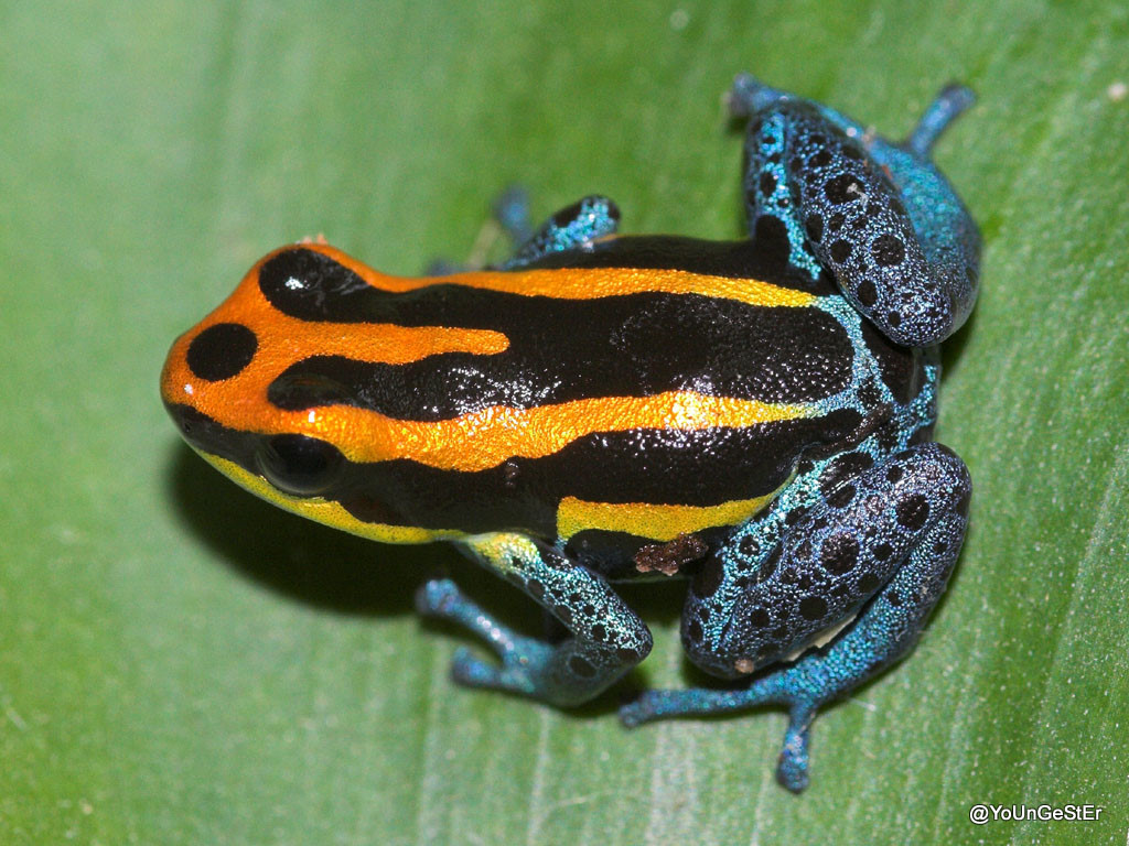
Ranitomeya amazonica 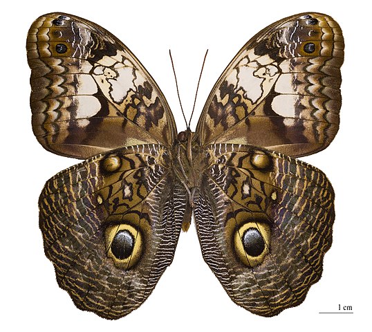
Caligo martia
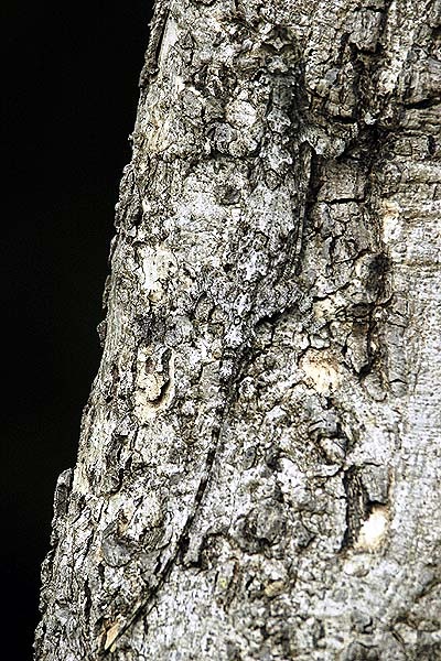
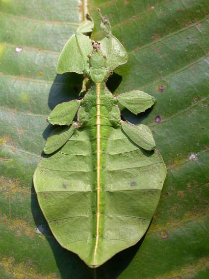
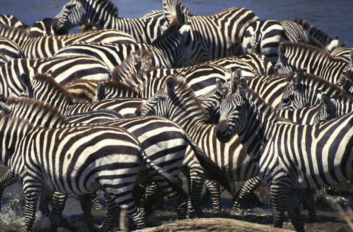
Another strategy is that of cryptic coloration, otherwise known as camouflage. This type of defense makes organisms hard to see, allow them to better hide from predators. For example, lizards that best match the color of the tree bark on which they live are less likely to be eaten by visual predators, and insects shaped like leaves can more successfully blend into the plants on which they live. Disruptive camouflage – like the stripes on a zebra – can break up the silhouette of these animals, especially when they are in a herd, making it more difficult for predators to recognize them.