Student landing page: https://wendystjohn.summerlark.net/2021/02/24/131-deuterostomes/
LAB SAFETY SHEETS
Please make sure that ALL students have signed the lab safety sheet. Once it is complete, you can drop it in my box in the Biology Office.
EXAMINATION OF SEA URCHINS
Begin this activity by showing students this video. If you want, play it once at normal speed, and then adjust the “Settings” to view it at .25 speed:
They will now be able to remove a sea urchin from the marine tanks to view it more closely under a dissecting scope. Before removing an urchin from the tank to observe it, ask them to place a small piece of seaweed on its aboral (opposite the mouth) surface (the small plate on the top of the urchin). Then they should observe the behavior.
IMPORTANT:
- Before handling the urchins and sea stars, it is necessary to rinse the hands in fresh tap water, to remove any traces of hand sanitizer, lotion, etc.
- When placing an urchin in a finger bowl, make sure there is a small amount of seawater in the bowl.
- Students should only have the urchins out of the tank while viewing them, and then they should be returned to the tank,
- Do not leave the urchins under the dissecting scope lights when they are not being actively viewed.
- When returning urchins to the tank, make sure to put the seawater back in the tank, as well. If an urchin has attached itself to the finger bowl, you can place the entire bowl in the tank, and then remove it in a few minutes after the urchin has moved out of the bowl on its own.
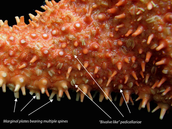
They can use a dissecting scope to identify the spines, tube feet, and pedicellariae. They are also being asked to sketch a few pedicellaraie, with a representative sample of the various kinds they observe.
Place a sea urchin in a finger bowl with a small amount of seawater. Identify the spines, tube feet and pedicellariae. Use the dissecting scope to examine the pedicellariae more closely.
- Before removing an urchin from the tank to observe it, place a small piece of seaweed on its aboral surface. Describe what happens to it.
- Observations will vary
- Place a sea urchin in a finger bowl with a small amount of seawater. Identify the spines, tube feet and pedicellariae. Use the dissecting scope to examine the pedicellariae more closely. Are they all the identical?
- Not usually – they may be of different lengths or have 2 or 3 “claws.”
- Look at both the oral and aboral surfaces. Draw the different kinds of pedicellariae you observe.
- What happens if you touch the urchin with a sea star?
- Observations will vary
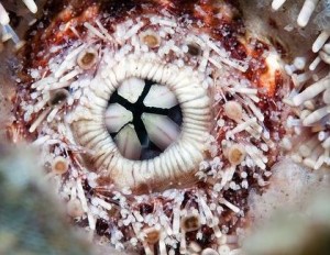
- Turn the sea urchin so that its oral side is facing upwards. Examine the mouth area using the dissecting microscope. Look for the 5 teeth that connect to the internal jaws called Aristotle’s lantern. Compare what you see on the live animal to the Aristotle’s lantern on display. Draw both.
- The ossicles that make up the sea urchin’s test are internal, but surround all the visceral organs. Arthropods molt their exoskeleton to grow. When sea urchins grow they need to increase the size of their test to accommodate their increasing viscera. Since they don’t molt, and their test surrounds their internal organs, how do you think they manage to make their test grow larger as they grow? Where do they add new material?
- These calcareous plates grow by apposition: they add material or erode it only at the margins of the individual plates. (bone also grows appositionally).
EXTERNAL EXAMINATION OF SEA STARS
There are several sea stars available for them to look at. In the tank, see if any of them are feeding. If they are, look for their everted stomach when they are being removed from the tank. Sea stars eat by everting their stomachs onto or into their prey, and then digesting and absorbing them externally.
Show them this video before they put specimens in finger bowls:
- Place the seastar in a bowl with some seawater from the tank. Look for the madreporite in the central region (central disc) on the aboral surface. This is the entrance to the water vascular system. The fleshy projections along the surface of the body are dermal branchae and function in gas exchange.
- Can you see any pedicellariae? Are they the same or different from the ones on the sea urchin? Draw all the structures you see on the aboral surface of your specimen under the dissecting microscope.
- Turn the star over and examine the oral surface. Draw an oral view of the animal. Identify the ambulacral groove and the tube feet.

SEA STAR DISSECTION
Show them this video that shows a starfish dissection.
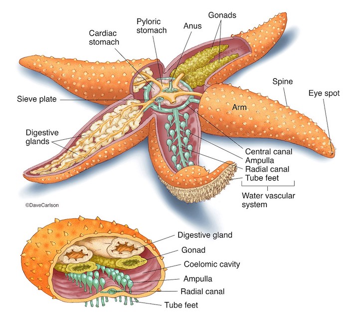
There is a diagram in the worksheet for them to label:
- Coelom—the space containing the internal organs/viscera
- Stomach—5-‐lobed, sac-‐like structure dorsal to the mouth in central disc region, connects to caeca
- Digestive glands—pair in each arm connect to stomach. Also known as hepatic caeca.
- Gonads—found under the caeca in each arm. Testes or ovaries (sexes separate in echinoderms)
- Water vascular system: includes
- Stone canal: connects madreporite to ring canal
- Ring canal: hard, circular tube-‐like structure, around mouth
- Tiedemann bodies: swellings (9) along ring canal
- Radial canal: from ring canal down each arm, connects water vascular system to ampullae
- Ampullae: spherical “bulbs” connect to tube feet
Acivity 1d. Display of additional echinoderms. There may also be examples of holothuroidians (not always available) and ophiuroids on display.
Draw a sea cucumber (optional) and a brittle or basket star. Which one of these is the holothuroidian? Can you locate tube feet on each of them? What about ambulacral grooves? Can you discern pentamerous symmetry in these taxa?
SHAPE OF LIFE: ECHINODERMS
Show students the video at the link below. Please make sure that Closed Captions are turned on. You can also provide the link to students if it means they are better able to view the transcript. While they are watching, they have questions in the worksheet to answer.
https://video.lanecc.edu/media/The+Shape+of+Life+Vol.+7+-+Ultimate+Animal/0_ad0xz0ze/84523512
- What allows seastars to hold their position effortlessly for hours?
- A special protein allows them to lock their arms in position effortlessly for hours, something our muscles could never do
- How fast can an urchin eat seaweed?
- Every hour, a lettuce-sized piece of kelp can disappear down those star-shaped jaws. When urchin populations are high, the persistent nibbling of their armies can mow down an entire kelp forest within a matter of months.
- How do soft-‐bodied sea cucumbers defend themselves from predators?
- The truth is they have few predators. Some simply taste bad. Bold patterns help advertise the noxious toxins which lace their bodies. Unlike urchins, sea cucumbers have no pointy quills to protect them, just fleshy knobs that cover their skin. Still, when provoked, some cucumbers can unleash an entirely unique defense. Until it was disturbed, this weapon was actually part of the sea cucumber’s breathing apparatus. Ejected from its anus, these thin, white strands swell to 20 times their original size, packing a potent mix of poison and glue.
- What do brittle stars eat?
- Plankton
- Can seastars see? If yes: Can they see light? Can they form an image?
- The tip of each arm is also equipped with a light-sensing organ seen here as a red dot. This receptor cannot form images, but it can sense light and darkness
- Describe how a seastar eats a bivalve.
- Settling on its victim, the sea star hunkers down and begins its attack. The first wave of the assault begins with the tubed feet which use hydraulic pressure to pry open any gaps in the mussel’s shell. Next, the sea star deploys its most unusual weapon – its stomach which moves out of its body to digest its prey alive. Even in time-lapse, it is hard to see the sea star’s attack. But a miniature camera tucked within the mussel’s shell gives us the first look ever at the carnage that unfolds here every day. Once the tubed feet have physically breached the mussel’s defensive line, the sea star’s translucent stomach begins the final assault. The animal actually pushes its stomach inside the mussel’s shell unfolding like a fatal flower. The stomach unleashes a volley of chemical weapons, digestive juices that dissolve the mussel’s soft pink flesh. All that’s left is a nutrient-rich soup, a broth, that’s quickly absorbed by the sea star. Having assimilated the mussel, the sea star’s stomach pulls away and the animal moves on leaving behind an empty shell.