During today’s lab, you will complete three activities:
- Mitosis Mover
- Introduction to Microscopy
- Onion Root Mitosis
You will also complete the Henrietta Lacks activity for homework (see the Canvas page for details).
Worksheet: https://docs.google.com/document/d/1rSE_ngUkK-vtUt0RNPZyQHZUGP9SG8yhd_95Byiq83E/copy
Activity – Mitosis Mover!

In this activity, you will work through an interactive website where you will learn about the cell cycle, including the process of mitosis:
https://biomanbio.com/HTML5GamesandLabs/Genegames/mitosismoverpage.html
Once you “Start a New Game,” simply follow the instructions. All of your answers will be saved, and at the end you will print out a screenshot to document your progress. Along the way, keep an eye out for the answers to the questions below.
Interphase:
- What must happen to the DNA before mitosis can occur?
Prophase:
- What happens to the chromosomes during this phase
- What is the name for each half of the chromosome?
- What happens to the nuclear membrane?
Metaphase:
- Describe what happens to the chromosomes during metaphase.
Anaphase:
- Which structures are separated from one another during anaphase?
- Where do these structures end up?
Telophase:
- What forms around each set of chromosomes during telophase?
Cytokinesis:
- What happens during this phase of mitosis
- In the space below, insert a screenshot of the resulting daughter cells:
Follow up questions:
- 7. How many copies of each chromosome were present in the cell before S phase?
- 8. How many copies of each chromosome are present in each of the resulting daughter cells?
- 9. What is the genetic relationship of the daughter cells to one another? To the parent cell?
- 10. In the space below, insert a screenshot of the stages of mitosis and cytokinesis after you have put them into the correct chronological order.
- 11. In the space below, insert a screenshot of your final Mitosis Mover Score.
Activity: Introduction to Microscopy
Working magnification
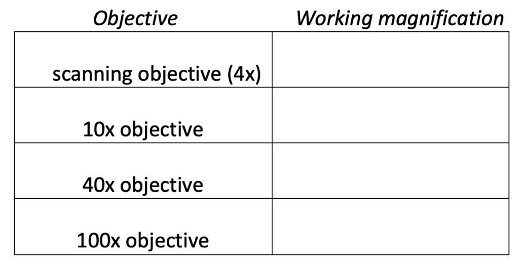
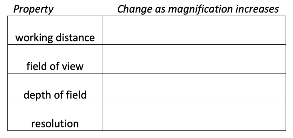
Working magnification (total magnification) is the product of the ocular magnification (usually 10x) and the objective lens magnification (ranging between 4x and 100x). The following terms refer to characteristics of a particular working magnification.
- Working distance is the distance between the focal plane (the plane in the specimen where you are focused) and the front of the lens.
- Field of view is the area in the focal plane that is visible through the objective.
- Depth of field is essentially the thickness of the focal plane, or more precisely, the range of depth in which the specimen is close enough to being in perfect focus.
- Resolution is the size of the smallest feature that can be distinguished.
Questions:
- Determine the working magnification for the following objectives:
2. How do the properties of working magnification change when you increase magnification?
Onion Root Tip Mitosis
Growth in an organism is carefully controlled by regulating the cell cycle. For example, in plants, the roots continue to grow as they search for water and nutrients. These regions of growth are good for studying the cell cycle because at any given time, you can find cells that are undergoing various stages of mitosis.
Today, we will examine cells in the tip of an onion root. In order to view them under a microscope, a thin slice of the root is placed onto a microscope slide and stained so the chromosomes will be visible. Although slicing the onion root captures many cells in different phases of the cell cycle, keep in mind that the cell cycle is a continuous process.
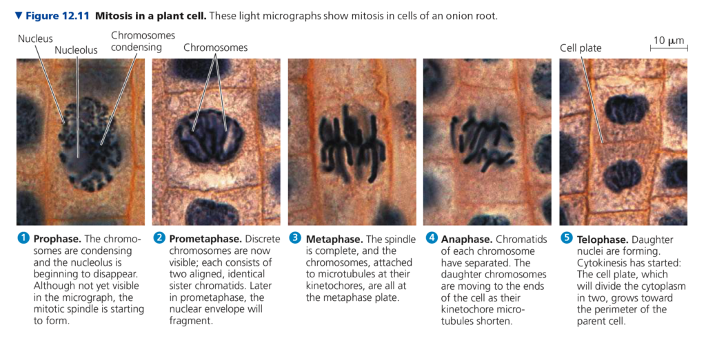
Activity: Mitosis in Onion Root Tips
1) Virtual Exercise
Work through the activity on the website below, where you will learn to identify the stages of mitosis in the cells at the tip of an onion root, and classify each cell based on what phase it is in.
http://www.biology.arizona.edu/cell_bio/activities/cell_cycle/cell_cycle.html
- As you classify the cells, keep a tally in the table below:
| Interphase | Prophase | Metaphase | Anaphase | Telophase/ Cytokinesis | Total | |
| Number of cells | 36 | |||||
| Percent of cells | 100% |
Count up the cells found in each phase and use those numbers to predict how much time a dividing cell spends in each phase. Divide the number of cells in each phase by the total number of cells classified (this gives you the percentage for each). Enter the percentages into the bottom row of the table above.
- In this virtual exercise, which stage of the cell cycle is the longest?
- Why do you think root tips might be a good choice for viewing cells in different stages of the cell cycle?
2) Onion Root Microscopy
Using the Virtual Microscope you explored in the activity earlier today, find the onion root tip slide. From the slide box, select “Plant Slides,” and then “Onion Root.” Once again, zoom in and focus on the slide until you are using the 40x objective lens. This gives you an idea what this would look like through the microscope, because of the limited view through this microscope, you have the option to assess the stages of mitosis using the photograph below.
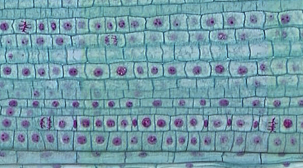
Work with a partner to categorize the stages of mitosis in the cells you see in the image, just as you did during the demonstration. If you can’t tell which stage of mitosis a cell is in, you can categorize it as being in Interphase.
In the space below, draw and label an example of each of the 5 cell phases:
| |
Now, count as many cells as you can, using the table below to tally the number of cells in each phase:
| Phase | Tally # of Cells | Total |
| Interphase | ||
| Prophase | ||
| Metaphase | ||
| Anaphase | ||
| Telophase/Cytokinesis |
Once you have counted the cells on the slide, visit the link below, which will take you to a Google Spreadsheet. Please note: You must MAKE A COPY OF THE SPREADSHEET! Do not request permission to edit! You must copy the file to your own Google Drive. (If every student tried to enter data into the same spreadsheet, it would get chaotic very quickly)!
https://docs.google.com/spreadsheets/d/1CijQY8l089f0pcQFnZzn84juwc7GHXXc8l7c4myxbcc/copy
Enter your tally data into the spreadsheet, on the tab labeled “Onion Root Microscopy.” This will generate a graph for you, of the time spent in each phase of mitosis.
- In the space below, recreate the graph from the spreadsheet:
- Did you have similar results across the two exercises? If not, why do you think this might be?