Before your lab section meets, please read through the information below and view all videos. If you have a computer or tablet that you can bring with you to lab, it might be helpful for you to be able to access this material during lab. Don’t forget to return to Canvas and take the weekly pre-lab quiz. NOTE: You do not need to print out a worksheet before lab. The worksheet will be provided to you by your lab instructor.
Humans have been familiar with macroscopic organisms (organisms big enough to see with the unaided eye) since before there was a written history. It is likely that most cultures distinguished between animals and land plants, probably including the macroscopic fungi as plants. Therefore, it became an interesting challenge to deal with the world of microorganisms once microscopes were developed a few centuries ago.
Many different naming schemes were used over the last couple of centuries, but it has become the most common practice to refer to eukaryotes that are not land plants, animals, or fungi as protists.
The word “protist” was first suggested by Ernst Haeckel in the late nineteenth century. It has been applied in many contexts and has been formally used to represent a kingdom-level taxon called Protista. However, many modern systematists (biologists who study the relationships among organisms) are beginning to shy away from the idea of formal ranks such as kingdom and phylum. Instead, they are naming taxa as groups of organisms thought to include all the descendants of a last common ancestor (monophyletic groups). During the past two decades, the field of molecular genetics has demonstrated that some protists are more related to animals, plants, or fungi than they are to other protists. Therefore, not including animals, plants and fungi make the kingdom Protista a paraphyletic group, or one that does not include all descendants of its common ancestor. For this reason, protist lineages originally classified into the kingdom Protista continue to be examined and debated. In the meantime, the term “protist” still is used informally to describe this tremendously diverse group of eukaryotes.
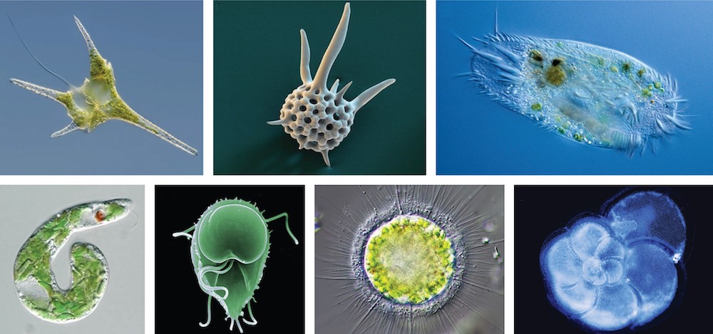
Most protists are microscopic, unicellular organisms that are abundant in soil, freshwater, brackish, and marine environments. They are also common in the digestive tracts of animals and in the vascular tissues of plants. Others invade the cells of other protists, animals, and plants. Not all protists are microscopic. Some have huge, macroscopic cells, such as the plasmodia (giant amoebae) of myxomycete slime molds or the marine green alga Caulerpa, which can have single cells that can be several meters in size. Some protists are multicellular, such as the red, green, and brown seaweeds. It is among the protists that one finds the wealth of ways that organisms can grow.
There are over 100,000 described living species of protists, and it is unclear how many undescribed species may exist. Since many protists live as commensals or parasites in other organisms and these relationships are often species-specific, there is a huge potential for protist diversity that matches the diversity of hosts. As the catchall term for eukaryotic organisms that are not animal, plant, or fungi, it is not surprising that very few characteristics are common to all protists.
Text adapted from “Biology” by Openstax College. Download for free at: http://cnx.org/content/col11448/latest/
Porifera
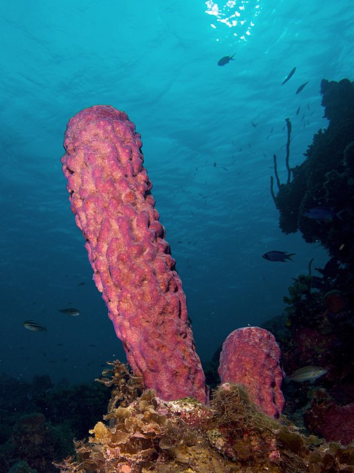
The sponges (phylum Porifera), jellies, hydroids, corals and sea anemones (phylum Cnidaria) are marine and freshwater organisms. These two groups differ from the rest of the Metazoa (multicellular animals) in substantial ways. Unlike many multicellular animals, the bodies of these organisms are not always tightly integrated into a single functional unit. They often grow into asymmetrical or irregular shapes (sponges) or as repeated modular units like plants (some hydroids, sea anemones, corals).
Many of these animals are sessile as adults, but have free-swimming larvae (or other life history stages) that aid in dispersal to new, suitable substrates for settlement, metamorphosis and growth into adult forms.
- Cellular level of organization
- No tissues
- Asymmetrical body form
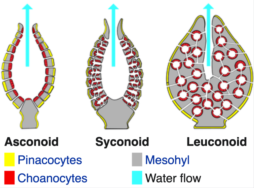
- Cavity consisting of a spongocoel lined with flagellated cells called choanocytes (or collar cells)
- Acellular internal support elements called spicules
The phylum Porifera (“pore bearers”) contains approximately 5,000 sponge species, classified into three groups based on the chemical composition and structure of the spicules that provide structural support for these animals. Three main body forms occur: Asconoid, Syconoid, and Leuconoid. The asconoid sponges are vase shaped, with the simplest arrangement of canals and are typically very small. The syconoid sponges are still vase shaped but they have a more complex network of canals. Both of these groups have a large central cavity (spongocoel) with a conspicuous osculum. The leuconoid sponges are most common and have a complex network of canals, tend to be larger with a globular or crustose morphology, and have several smaller oscula.
Preparing a Wet-Mount Slide
A wet-mount slide is when the sample is placed on the slide with a drop of water and covered with a coverslip, which holds it in place through surface tension. To make your own wet-mount slides:
- Use a pipette to extract a small amount of pond water from your sample. If there is any obvious algae or vegetation in the water, try and get a bit of it in the sample, as there is a good chance that there will be organisms associated with that material.
- Place a single drop of water at the center of your slide. (Do not use too much water or else your coverslip will float away and you won’t be able to see your sample.)
- Place a single drop of Proto-slo quieting solution on top of your water sample. This increases the viscosity of the water, slowing down the organisms for ease of viewing.
- Place a coverslip at a 45-degree angle with one edge touching the water and let go. The coverslip will fall over the sample and be held in place by surface tension. (This method should prevent any air bubbles under the coverslip, as these will affect your ability to view the sample.)
- Your slide is ready to be viewed.
To complete this activity, you will work with the wet-mount slides you prepared from pond water collected on your campus walk, and from the samples provided to you in the classroom. View the slide(s) under the microscope, and see if you can identify any organisms you see. Start with the identification guide in the worksheet, and if you need to do more investigation, the links below will take you to good online identification guides.
- https://microscope-microscope.org/pond-water-critters-protozoan-guide/
- http://www.microscopy-uk.org.uk/index.html?http://www.microscopy-uk.org.uk/pond/index.html
- You may also wish to do a Google search for additional information once you have a guess as to the ID
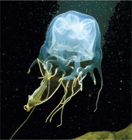
Phylum Cnidaria
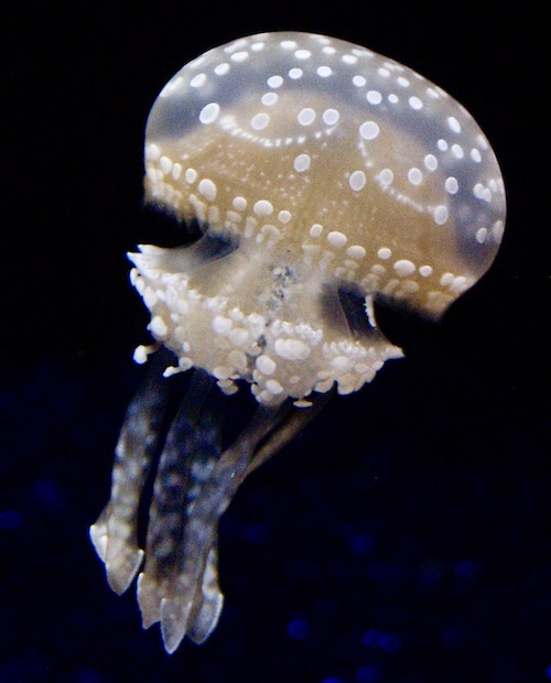
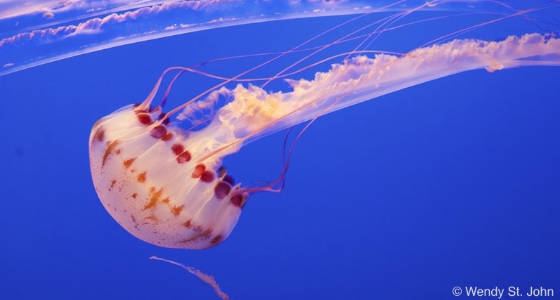
About 10,000 species of Cnidaria exist. All are aquatic and most are marine. These include the jellies, corals, hydroids and sea anemones. Cnidarians have two forms. Both show radial symmetry: the polyp (sessile) and the medusa (free-living). Cnidarians reproduce both sexually and asexually, and individuals of some species spend part of their lives as a polyp and part as a medusa. The sexual stages usually are dioecious (with female and male individuals). Most cnidarians are predators that feed on zooplankton, but a few eat larger organisms, including fish, mussels and urchins. They obtain prey using their cnidae, organelles unique to the phylum, that can harpoon, spear and paralyze their prey, or simply function like a ‘Velcro’ sticky trap (see lecture notes). We will introduce the four cnidarian classes, and investigate two of them more closely.
The classes differ in structure and in the relative dominance of the polyp versus medusa stage.
Characteristics:
- Radial symmetry
- Tissue level of organization
- Two germ layers (diploblastic -‐ endoderm and ectoderm)
- A middle layer of jelly-like mesoglea
- Gastrovascular cavity originating from the blastocoel
- Single opening for mouth and anus that is surrounded by tentacles
- Cnidocytes present for prey capture
- Sessile or free-‐floating
- Sexual and asexual reproduction
- Two main body forms
- Polyp: sessile hydroid form
- Medusa: floating jelly form
CLASS HYDROZOA-: both polyp and medusa present with polyp stage dominant, solitary or colonial, smaller medusae than Scyphozoa, polyps asexual, medusae sexual, freshwater or marine.
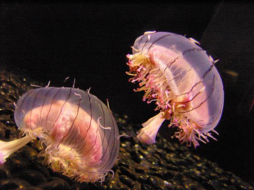
CLASS SCYPHOZOA: polyp stage reduced or absent; medusa stage dominant, polyps asexual, medusae sexual, medusae bell-shaped, marine.
CLASS ANTHOZOA: solitary or colonial, only polyps (sexual or asexual) in life cycle, no medusae, digestive cavity of polyp subdivided by septa, marine.
CLASS CUBOZOA: solitary, medusa stage only, cube body shape, 4 sets of tentacles, well developed eyes, highly venomous, about 20 species, marine.
Fungi
Although humans have used yeasts and mushrooms since prehistoric times, until recently, the biology of fungi was poorly understood. In fact, up until the mid-20th century, many scientists classified fungi as plants. Fungi, like plants, are mostly sessile and seemingly rooted in place. They possess a stem-like structure similar to plants, as well as having a root-like fungal mycelium in the soil. However, molecular biology analysis of the fungal genome demonstrates that fungi are more closely related to animals than they are to plant.
Based on fossil evidence, fungi have been found in the Denovian era, about 410 million years ago, although new findings may provide evidence for fungi as early as 900 million years ago. Fungi are heterotrophs, (organisms that eat other organisms to survive, rather than performing photosynthesis), absorbing nutrients through their cell walls. For this reason, they act as decomposers, helping to recycle nutrients by breaking down organic materials to simple molecules.
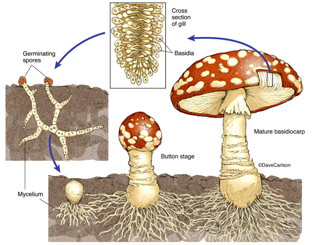
Some fungal organisms multiply only asexually, whereas others undergo both asexual reproduction and sexual reproduction with alternation of generations. Most fungi produce a large number of spores, which are haploid cells that can undergo mitosis to form multicellular, haploid individuals.
Fungi often interact with other organisms, forming beneficial or mutualistic associations. For example, most terrestrial plants form symbiotic relationships with fungi. The roots of the plant connect with the underground parts of the fungus, which form mycorrhizae, through which the fungus and plant exchange nutrients and water, greatly aiding the survival of both species. Lichens are another commonly seen mutualism, where fungi live symbiotically with a species of cyanobacteria or algae.
Fungi also cause serious infections in plants and animals. For example, Dutch elm disease, which is caused by the fungus Ophiostoma ulmi, is a particularly devastating type of fungal infestation that destroys many native species of elm (Ulmus sp.) by infecting the tree’s vascular system.
Text adapted from Open Stax: https://openstax.org/books/biology-2e/pages/1-introduction
Video Transcript:
00:00: Rod Nelson here. Now if you want to identify a mushroom the first thing that you’re probably going to do is you’re gonna get a nice book like this and you’re gonna go out into the woods.
00:07: You’re gonna look at your mushroom and try to figure out what you’re looking at. Now a book like this uses a lot of lingo so you’ve got to know what you’re looking at on the mushroom, so we’re gonna look at the basic parts of a mushroom right now.
00:18: I can’t get to all the parts and variations so we’re gonna look at just the classic capped mushroom and just look at the basics.
00:25: When you identify a flower, and this is what I’d used to do, I’d take a nice pretty colorful book like this and I flipped through. I’d find like the orange flower or the red flower try to figure out what I was looking at. I’d be like oh that looks about the same, then I flipped to the back, make sure I read up on it, and most of the time you’d be right but mushrooms are not like that.
00:42: They can vary in color, they can vary in size, they can vary in shape. It is amazing the diversity of one individual mushroom so you don’t really use, you know, the outward image to identify a mushroom.
00:57: So, what do you do? Well you look at key characteristics of that mushroom, so let’s start with the basic anatomy of a capped mushroom. I’m starting with that group because not only is it one of the more common ones you’ll see, it’s also the group where most of the poisonous and toxic mushrooms are found.
01:11: It might resemble this. On top you have what some people call a cap, also known as a pileus, a Stipe or stalk, and then at the bottom it’s actually connected to the actual mycelial threads – the meat of the fungi.
01:23: The gills – often called the lamellae – are under the cap and of course knowing the color of the spores which you can’t identify right away is important .
01:33: Sometimes along the stipe you have a ring known as the annulus and at the bottom sometimes you have a sac called the vulva. All of these are important to start your identifications in the wild.
01:42: Now, I gave you a few names for each of some of these basic parts of the mushroom. The reason for doing that is that even though some people argued that one is the more scientific way of saying it, in reality if you watch YouTube videos or you go to some of these books, different people use one or other of the names. The point is that you need to know a couple different names if you’re gonna take a book like this into the wild and start identifying your mushrooms.
02:07: All right, so that’s really the major stuff and I know you probably will be looking for more, so subscribe to this channel because I’m gonna get into all the nitty-gritties of all the different groups in future episodes
02:16: Alright see you then. This is the second mushroom video that I am releasing from my old YouTube channel because it is mushroom week and we’re leading up to the new year. A new year of content for untamed science. We are really excited about getting all my interests into this one channel. Now two links to note. Jonas and I make these videos here and we teach you how to do it, and this is my new Patreon site where you can support what I do and see fun behind-the-scenes stuff, and they know I have other perks in there. Alright see you tomorrow.