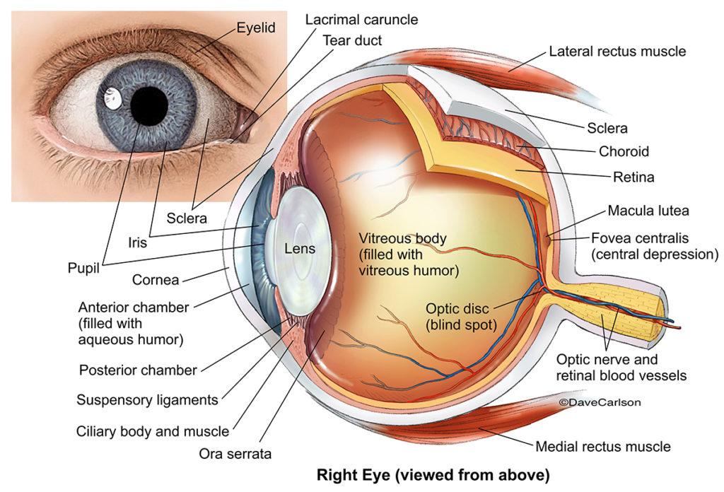
Rods and cones
The retina is a thin layer of neural tissue lining the back inside of the eyeball; this tissue contains the receptors for vision that are called “rods” and “cones.” Cones are color sensitive and work best in bright light. Rods are more light sensitive and work better in dim light but are not sensitive to color, so they are important to night vision. Cones are most densely concentrated in the center of the retina, while rods are more concentrated around the periphery of the retina.
Experiment 1
Sit in the dark for a few minutes to let your eyes adjust. In near darkness, stare directly at an object and attempt to make out the details.
Now turn your head away slightly to look at it using your peripheral vision (from the side). Can you make it out more clearly? How does this relate to rods and cones? Try this trick while stargazing and you’ll see more!
Experiment 2
Lay several colored objects in front of you, and very slowly bring up the lights from full darkness. Why can you see but not easily identify colors in dim light? How does this relate to rods and cones?
The Blind Spot
The retina is like a projection screen on which the image is focused by the lens of your eye. Within the retina however, there is a small area where the optic nerve connects to the eye. There are no light sensitive cells in this area, which is called the blind spot. No visual information is sent to the brain from the part of your field of vision projected onto the blind spot. One interesting bit of trivia is that the cephalopod eye (found in the invertebrate molluscs like octopus and squid) evolved independently from the vertebrate eye and, because of a different internal design, lacks a blind spot.
Experiment 1
Hold your left hand over your left eye while staring directly at the X with your right eye, and slowly approach the screen until the black spot disappears (about a foot away from the screen). You have found your blind spot. With both eyes open, why can’t you find your blind spot?

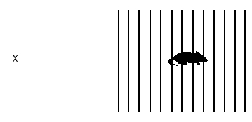
Experiment 2
Now repeat this procedure while looking at the “X.” When the mouse disappears, what happens to the bars of the cage? How can you “see” something in what you know is your blind spot? Why do you see the bars but not the mouse? Note: If you have trouble finding your blind spot, remember to move your head very slowly towards the screen with your left eye covered.
In some cases, as with the blind spot experiment, the brain fills in missing information to compensate for the “missing data” coming from the blind spot region, but it can only fill in a regular pattern by sampling the area around the missing data. In other cases, perception (which occurs in the brain) can be fooled by what the eye sees accurately. In each of these examples, try to identify why your perception is being fooled. Note that an image that comes to the eye is projected upside down onto the retina. How is it, then, that we see things “right side up”?
Afterimages
A change in sensitivity limited to a restricted region of the retina is called local adaptation. This can result in the production of afterimages. With one eye covered, fixate on the white dot in the center of the “peace symbol” on the left for about 30 seconds. Then fixate on the black dot in the middle of the circle to the right. Describe what you see in the white circle. How has the sensitivity of your retina changed after staring at the dark geometric shape? How long do you continue to see the afterimage?

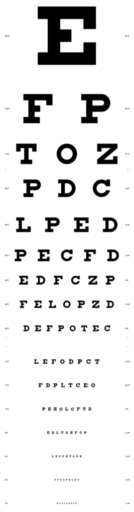
Vision Test
20:20 vision means that at a distance of 20 feet, you can distinguish the letters on a chart as well as the average person can at 20 feet. A person with 20:100 vision is very nearsighted because what they can only see at 20 feet, an average person can see at 100 feet. A person with 30:20 vision has better than average “visual acuity” because they can see at 30 feet what the average person can only see at 20. Sometimes these “acuity ratios” are divided through to give a single number. In that case, a number greater than 1 means above average visual acuity while a 1 is average and a number less than 1 indicates below average visual acuity.
Adjust the image below so that the big E is 3.5 inches tall. Then, scroll down a few lines, and move 20 feet away from your computer screen and attempt to read the chart If you can accurately read the line marked 20 ft. from 20 ft. away, you have 20:20 vision. (You will need to scroll down a few times to view the entire chart). If you wear glasses, take the test with and without them and compare results.
Color Blindness
Due to a mutation on the X chromosome, males are more likely to express color blindness than females since females (who are XX) have a “backup” X and males (who are XY) don’t. About 5% of males have some degree of red-green color blindness. Do you know any colorblind men? Do you know any colorblind women? On the following test chart, everyone can easily distinguish the orange circle but people with red-green colorblindness will have difficulty identifying the red star. How did you do?

If you are interested in taking another colorblindness test, you’ll find one here: https://www.color-blindness.com/rgb-anomaloscope-color-blindness-test/