Worksheet:
Sea Urchin Anatomy
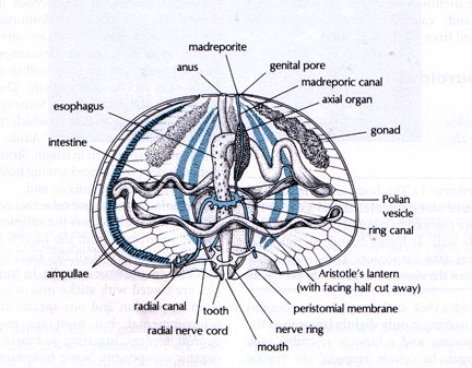
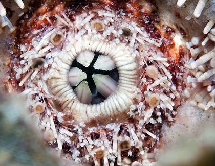
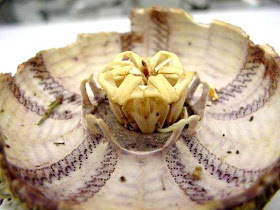
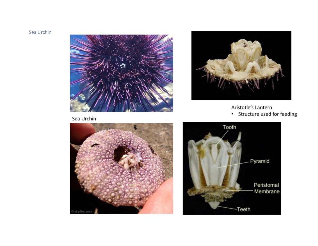
The feeding structure on a sea urchin is called Aristotle’s Lantern, due to its unusual shape. Using the materials below (and following the drawing guidelines we presented earlier this semester), do a sketch of Aristotle’s lantern, labeling as many structures as you can. Scan and insert your sketch into the worksheet.
Sea Star Anatomy
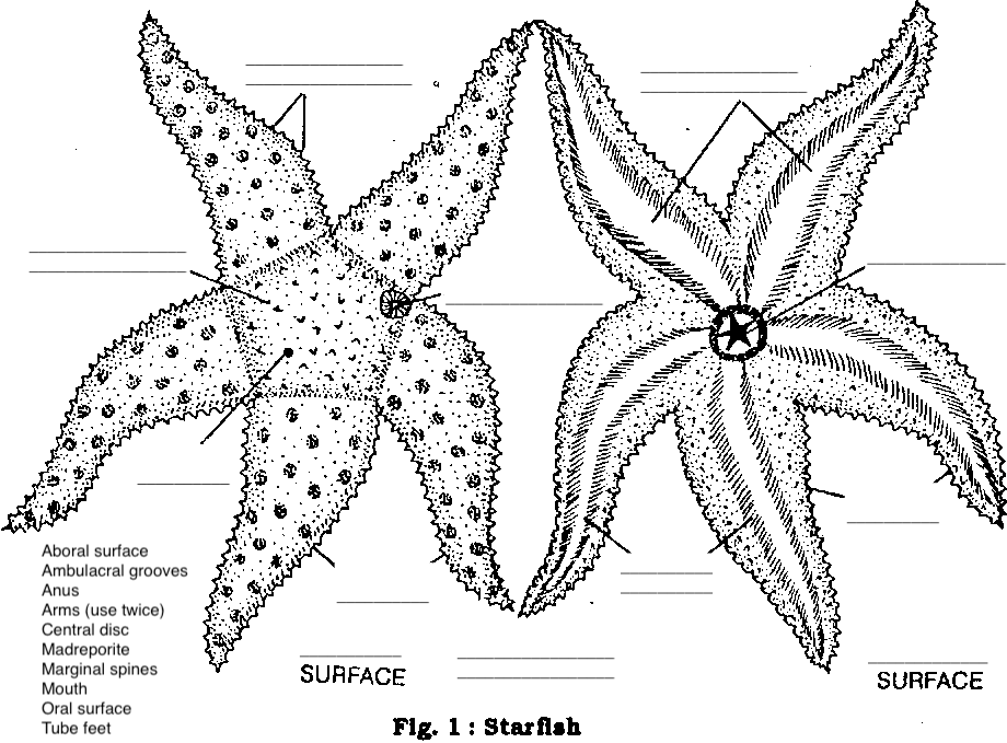
- Coelom—the space containing the internal organs/viscera
- Stomach—5-‐lobed, sac-‐like structure dorsal to the mouth in central disc region, connects to caeca
- Digestive glands—pair in each arm connect to stomach. Also known as hepatic caeca.
- Gonads—found under the caeca in each arm. Testes or ovaries (sexes separate in echinoderms)
- Water vascular system: includes
- Stone canal: connects madreporite to ring canal
- Ring canal: hard, circular tube-‐like structure, around mouth
- Tiedemann bodies: swellings (9) along ring canal
- Radial canal: from ring canal down each arm, connects water vascular system to ampullae
- Ampullae: spherical “bulbs” connect to tube feet
SHAPE OF LIFE: ECHINODERMS
Watch the video at the link below, and answer the questions in your worksheet.
https://video.lanecc.edu/media/The+Shape+of+Life+Vol.+7+-+Ultimate+Animal/0_ad0xz0ze/84523512
- What allows seastars to hold their position effortlessly for hours?
- How fast can an urchin eat seaweed?
- How do soft-‐bodied sea cucumbers defend themselves from predators?
- What do brittle stars eat?
- Can seastars see?
- If yes: Can they see light?
- Can they form an image?
- Describe how a seastar eats a bivalve.
